| Major Groups > Cup Fungi / Jelly Fungi > Urnula padeniana |

|
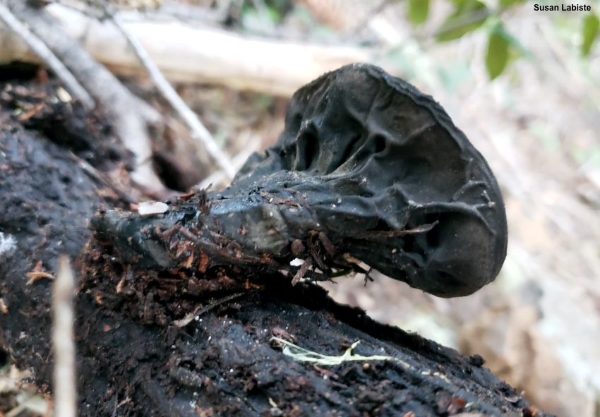
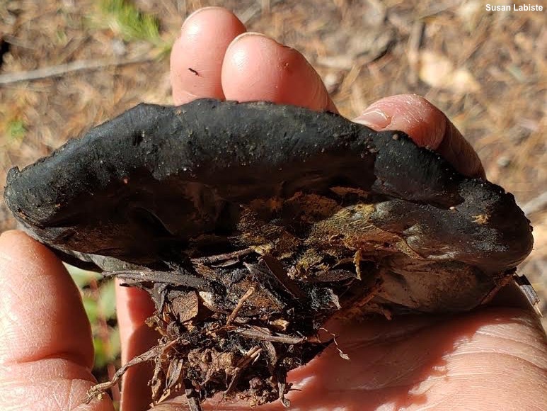
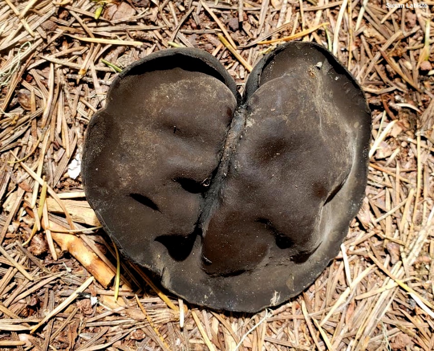
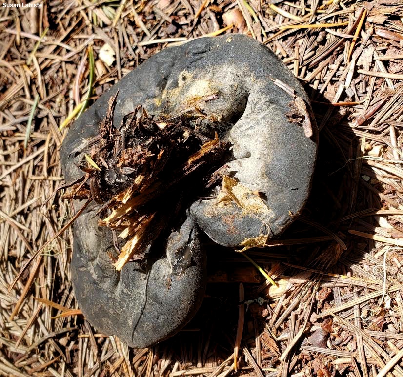
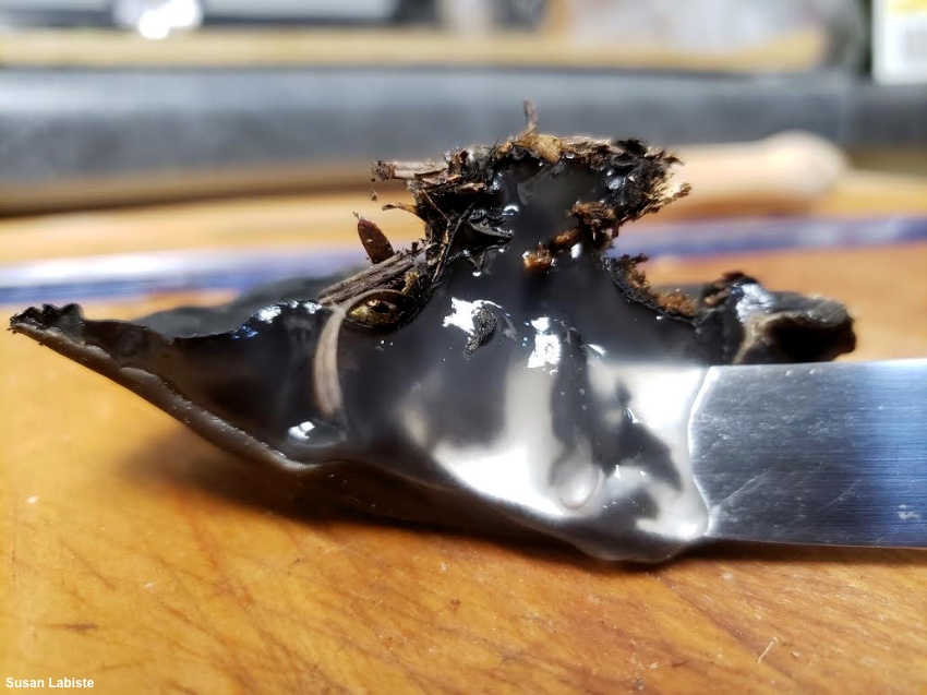
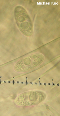
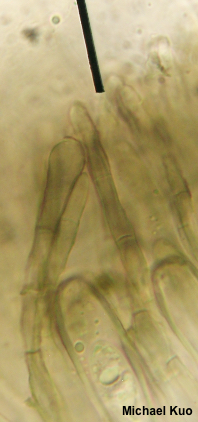
Urnula padeniana [ Ascomycota > Pezizales > Sarcosomataceae > Urnula . . . ] by Michael Kuo One of the Pacific Northwest's "snowbank fungi," Urnula padeniana appears on the wood or duff of conifers in the weeks after snowmelt in montane areas. It is an almost unmistakeable species: a large cup fungus with dark brown to black surfaces, a wrinkled stem or pseudostem, and (this is the clincher), an interior composed of thick, gelatinous, nearly hyaline flesh. I said "almost" unmistakeable because there is a similar fungus, also from Pacific Northwest snowmelt. Pseudosarcosoma latahense is bit smaller, has a purple-brown upper surface, and features slightly different spores that do not have oil droplets. Other potential look-alikes do not appear in Pacific Northwest snowmelt, and include Urnula mexicana (see the next paragraph), ranging from Mexico to California and appearing in fall, as well as Europe and northeastern North America's Sarcosoma globosum, which is rounder, browner, and lacks a stem. This fungus was previously called Sarcosoma mexicanum (or, with a Latin agreement error, "Sarcosoma mexicana") but Carbone and collaborators (2013b) discovered that two species were going under this name, and that they were actually quite distinct. Study of the type collection of Bulgaria mexicana revealed it to be a smaller, thin-fleshed, less gelatinous species with differently shaped spores. This meant that the fungus described here required a new name; the authors chose "padeniana" to honor J. W. Paden, a mycologist who had previously worked with Sarcosoma species in North America. In a separate paper (2013a) the researchers provided molecular support for placement of these mushrooms in the genus Urnula. Thanks to Susan Labiste for documenting, collecting, and preserving Urnula padeniana for study; her collections are deposited in The Herbarium of Michael Kuo. Description: Ecology: Probably saprobic; growing alone, scattered, or gregariously on the deadwood or duff on conifers; late winter through early summer, usually appearing within a few weeks of snowmelt; distributed in the Pacific Northwest. The illustrated and described collections are from Oregon. Fruiting Body: 5–10 cm across; 3–5 cm high; shaped more or less like a top, with a cup- or platter-like upper surface, round or slightly lobed in outline—and a narrowing stem portion; becoming more flattened overall with age. Fertile (upper, or inner) surface: Dark brown to black; smooth and bald. Sterile (lower, or outer) surface: Black; wrinkled; finely roughened, or velvety. Stem or Pseudostem: Poorly defined at apex; 2–5 cm high; 2–3 cm wide; tapering to base; black; wrinkled or even ribbed; finely velvety. Flesh: When fresh thick and gelatinous; nearly colorless; with age shriveling and rubbery. Odor: Not distinctive. Microscopic Features: Spores 20–28 x 10–12 µm; ellipsoid or very slightly fusiform; smooth; walls about 1 µm thick; hyaline in KOH and multiguttulate with many small guttules and usually, 2 or more larger guttules; in Melzer's with faintly brownish walls and no guttules. Asci 8-spored; cylindric; hyaline in KOH; tips not bluing in Melzer's. Paraphyses 3–5 µm wide; about the same length as the asci; filiform; frequently septate; terminal cells cylindric with rounded, slightly pinched, or subclavate apices; brown-walled in KOH; brown and somewhat agglutinated en masse. Context composed of gelatinizing hyphae 2.5–7.5 µm wide, smooth, hyaline in KOH, occasionally septate. Excipular surface elements cylindric; 5–7.5 µm wide; septate; brown in KOH; smooth or a little encrusted; occasionally branching and/or developing lobes or nodules. REFERENCES: M. Carbone et al., 2013. (Paden & Tylutki, 1969; Smith, 1975; Arora, 1986; Phillips, 1991; Trudell & Ammirati, 2009; Carbone et al., 2013a; Carbone et al., 2013b; Beug et al., 2014; Desjardin, Wood & Stevens, 2015.) Herb. Kuo 04292001, 05072001. This site contains no information about the edibility or toxicity of mushrooms. |
© MushroomExpert.Com |
|
Cite this page as: Kuo, M. (2020, May). Urnula padeniana. Retrieved from the MushroomExpert.Com Web site: http://www.mushroomexpert.com/urnula_padeniana.html |