Major Groups > Cup Fungi

MushroomExpert.Com

Cup Fungi  

[ Ascomycota . . . ]

by Michael Kuo

"Cup fungi" is not a very scientific term, but it holds together many mushrooms that are shaped more or less like cups, saucers, or goblets. In fact the cup-shaped mushrooms are very diverse, comprising several different families and genera in the Ascomycota.

Some cups are easily identified, but many are extremely difficult, requiring microscopic analysis; brownish, stemless cups are especially frustrating. Fortunately most cup fungi are fairly simple in construction, which means that there are not a lot of features to assess—even under the microscope.

Macroscopic features you will need to observe in order to attempt identifying cup fungi include the upper surface of the cup (the spore-producing "hymenium," in Mycologese), the margin (use a hand lens; you are looking for tiny hairs or pustules), the undersurface (the sterile "excipular surface"), the stem or pseudo-stem (if present), and the flesh.

Microscopic features usually involve the hymenium, so a scalp section from the cup's upper surface, crushed under your coverslip, will generally suffice. Morphology of the spores, paraphyses, and (sometimes) the asci should all be studied. (One frustrating hurdle in the study and identification of cup fungi involves the fact that immature specimens are often collected, making study of the spores impossible.) Ideally you should mount your sections in 2% KOH and in Melzer's reagent, since the reaction of the ascus tips to Melzer's (bluing or not bluing) can help greatly in figuring out which genus your cup fungus belongs to.

Recent investigations (including Tedersoo and collaborators, 2006) have suggested the possibility that many cup fungi we previously assumed to be saprobic decomposers of forest litter are actually mycorrhizal symbionts with trees. Further studies will, hopefully, discover more precisely which cup fungi are mycorrhizal, and whether they are always mycorrhizal or, like the morels, "facultatively" mycorrhizal.


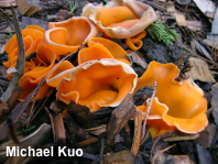
Aleuria aurantia

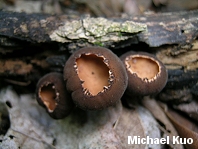
Galiella rufa

Peziza succosa


Partial, In-Progress Key to North American Cup Fungi  


NOTE: The key below delays using microscopic features for as long as possible—but does, inevitably, resort to them when the microscopic piper must be paid. Many places in the key are still undeveloped; I apologize to readers whose cup fungi are not yet included.


1.Cup initially (American) football-shaped and closed; later splitting into star-like rays; found in Texas, associated with stumps of cedar elm.

1.Cup not as above; range and ecology varying.
2


2.Cups when young growing partially underground (usually in clusters), the margin later peeling back in vaguely star-like rays; inner surface pale lilac to purplish when fresh; common in the Rocky Mountains and western North America, more rare in the east.

2.Not completely as above.
3


3.Growing in burned areas (burned forests, camp-fire pits, and so on).
4

3.Not growing in burned areas.
5


4.This portion of the key is not yet developed. For a few commonly collected species, see Geopyxis carbonaria, Peziza violacea, Rhizina undulata, and Sphaerosporella brunnea.


5.Margin of cup with tiny hairs, reminiscent of eyelashes or fringe (a hand lens may be required)—and/or undersurface of cup hairy (compare with option below).
6

5.Margin of cup without eyelashes or fringe; undersurface of cup bald, velvety, granular, finely fuzzy, scurfy, pustulate (and so on) but not hairy.
16


6.Upper surface whitish to creamy or pale bluish; undersurface with brown hairs that contrast with the paler surface underneath.
7

6.Upper surface dull to bright yellow, brown, orangish brown, red, or orange; hairs on undersurface variously colored.
9


7.Cup 2–7 cm across at maturity; usually with a ribbed pseudo-stem (often submerged in the ground) measuring up to about 2 x 2 cm; spores 25–30 µm long.

7.Cup 1–3 cm across at maturity; with or without a pseudostem; spores variously sized.
8


8.Stem absent; undersurface densely hairy, appearing more or less brown from the hairs; spores 20–25 µm long.

8.Pseudo-stem usually present (often submerged in the ground); undersurface sparsely hairy, appearing pale; spores 30–45 µm long.
Jafnea fusicarpa
(See Jafnea semitosta)


9.Cup goblet-shaped, fringed with prominent tufts of white hairs, about a centimeter across when mature; stem long and well developed.

9.Not as above.
10


10.Cup 3–7 cm across; upper surface dull to bright yellow or occasionally orangish yellow; outer/under surface dark brown to nearly black, hairy to woolly.

10.Cup well under 3 cm across; upper surface red to orange; outer/under surface varying.
11


11.Mature spores with oil droplets.
12

11.Mature spores without oil droplets.
14


12.Spores round; upper surface brown to orangish brown or brownish orange.

12.Spores ellipsoid; upper surface orange to red.
13


13.Cups to 3 mm across; mature spores smooth.

13.Cups to 15 mm across; mature spores with prominent warts.


14.Hairs pale yellowish or, at the most, light brown; growing on dung or occasionally on plant debris or soil.
Cheilymenia theleboloides

14.Hairs dark brown; growing only on dung.
15


15.Mature cup usually over half a centimeter across; hairs not branching (under the microscope).
Cheilymenia coprinaria

15.Mature cup usually less than half a centimeter across; hairs along the margin not branching, but hairs farther down the cup's outer surface branching.


16.Cups often clustered, arising from a shared, rudimentary stem structure; growing on the deadwood of hardwoods; cups up to about 3 cm across, often with a stretched appearance; paraphyses distinctively lanceolate and septate.

16.Cups not clustered and sharing a stem structure; growing on wood or not; variously sized and shaped; paraphyses not as above.
17


17.Cup fairly large (3–15 cm across at maturity) and fleshy; flesh gelatinous and thick (slice the mushroom open); outer surface brown to black; inner/upper surface ranging from brown to dull orange or dull yellowish.
18

17.Cup variously sized; flesh thin and brittle—or, if fleshy, flesh not gelatinous; surfaces variously colored.
21


18.Distributed from the Rocky Mountains westward.
19

18.Distributed from the Great Plains eastward.
20


19.Cups 5–15+ cm across at maturity; upper surface black to dark brown throughout development; most spores with several large oil droplets when mature.

19.Cups 2.5–5+ cm across at maturity; upper surface purple when young, becoming black; most spores lacking oil droplets when mature.
Pseudosarcosoma latahense


20.Found under conifers in northeastern North America; fruiting bodies at first nearly round, with an "eyeball" appearance, becoming squatty and lacking a stem; upper surface brown to blackish.
Sarcosoma globosum

20.Found under hardwoods, widely distributed east of the Great Plains; fruiting bodies with a stem or pseudostem throughout development; upper surface orangish to brownish orange.


21.Cup with a well developed stem that is fairly long in proportion to the cup (not stubby or rudimentary).
22

21.Cup without a stem, or with a stubby or rudimentary stem.
34


22.Growing from sticks or (sometimes buried) woody debris in spring in eastern North America; goblet-shaped when young; inner/upper surface black; outer/under surface brown to black, usually scaly; stem black, tapered to base.

22.Not completely as above.
23


23.Stem prominently ribbed; cup yellow brown to brown, grayish brown, or gray.
24

23.Stem not ribbed; cup variously colored.
26


24.Ribs not extending onto the undersurface of the cap.

24.Ribs, by maturity, extending onto the undersurface of the cup.
25


25.Cup yellow-brown; ribs with sharp edges.

25.Cup gray to grayish brown; ribs with blunt edges.
Helvella costifera
(= H. griseoalba)


26.Cup tiny and bright red; growing from sticks or buried woody debris in hardwood forests east of the Rocky Mountains.
27

26.Not completely as above.
28


27.Mushroom goblet-shaped; undersurface covered with long, whitish hairs.

27.Mushroom saucer-shaped or cup-shaped; outer surface not hairy.


28.Cups minute (4 mm wide or less); growing on hickory shells, acorns, and other "nutty" debris; uniformly whitish to pale yellow.

28.Not completely as above.
29


29.Cups bright orange; spores prominently reticulate; fairly common on the West Coast but rare elsewhere in North America.

29.Cup not orange; stem variously colored; variously distributed.
30


30.Cups darkly colored (very dark brown to black).
31

30.Cups paler than above (tan, grayish brown, brown, or gray).
32


31.Widely distributed in montane and northern North America; growing terrestrially in woods; spores with one large oil droplet.

31.Apparently limited to western North America; growing from woody debris under conifers, often in spring, near melting snowbanks; spores without oil droplets.
Plectania nannfeldtii


32.Mature spores spindle-shaped.

32.Mature spores elliptical.
33


33.Stem whitish, stout (about as long as the cup is wide, or shorter); cup pale to medium grayish brown.

33.Stem brownish to brown, not stout (usually longer than the cup is wide at maturity); cup medium to dark brown.
Helvella chinensis
(= H. villosa, H. pallidula)


34.Growing on wood. Note: Only some of the larger (over 1 cm) species are treated here. If your tiny, wood-loving cup is bright orange, see Lachnellula subtilissima. However, there are dozens and dozens of tiny (under 1 cm across), wood-inhabiting, cup-like species; see the sources by Dennis and by Breitenbach & Kränzlin, below, for an introduction.
35

34.Growing on the ground (if "the ground" is actually your carpet or flooring, see Peziza domiciliana).
59


35.Cup goblet-shaped, 2–5 cm across; flesh thick and gelatinous; upper surface drab orangish or reddish; outer surface dark brown or black; spores warted; growing in clusters on hardwood sticks and logs east of the Rocky Mountains.

35.Not completely as above.
36


36.Broadly attached to the wood so that only the extreme margin can be lifted; flesh somewhat gelatinous or rubbery.
37

36.Attached to the wood centrally, but not broadly; flesh fairly brittle.
40


37.Spores smooth.
38

37.Spores stippled or warted.
39


38.Mature cup less than 1 cm wide.
Pachyella babingtonii

38.Mature cup up to 5 cm wide.


39.Warts 1 µm or longer.
Pachyella adnata

39.Warts tiny (spores merely stippled in appearance).


40.Entire fruiting body blue to greenish blue; growing from greenish stained wood.
41

40.Not as above.
42


41.Spores 5–8 x 1–2.5 µm.

41.Spores 9–14 x 2–4 µm.
Chlorociboria aeruginosa


42.Upper surface red when fresh.
43

42.Upper surface otherwise colored.
47


43.Cup typically < 2 cm across; spores < 21 µm long; found east of the Rocky Mountains.

43.Cup larger; spores longer; variously distributed.
44


44.Found in tropical and subtropical regions (Florida, the U. S. Gulf Coast, Mexico); spores with longtudinal striations.

44.Variously distributed; spores not striated.
45


45.Found in the Pacific Northwest and California; spores usually unsheathed.

45.Found elsewhere; spores sheathed or not.
46


46.Mature spores with rounded ends; spores with several large (5–7 µ) oil droplets; spores when fresh and viewed in a water mount often encased by a full sheath; hairs on excipular surface not curling and twisted under the microscope.

46.At least some mature spores with flattened ends; spores with many oil droplets smaller than above; spores when fresh and viewed in a water mount lacking a full sheath but occasionally with "polar caps" (a sheathlike covering at each end); hairs on excipular surface curling and twisted under the microscope.


47.Cups bright yellow; under half a centimeter across.
48

47.Not completely as above.
51


48.Cup with a (proportionally) substantial stem; growing on small sticks or twigs.
49

48.Cup lacking a stem; growing on logs or stumps.
50


49.Growing on twigs of willows; spores 12.5–16 µm long; paraphyses not septate.
Hymenoscyphus conscriptum

49.Growing on twigs of other hardwoods; spores 16–24 µm long; paraphyses septate.


50.Cup 1–3 mm across; paraphyses with hyaline contents.

50.Cup 0.5–1.5 mm across; paraphyses filled with yellow droplets.
Bisporella sulfurina


51.Tips of asci bluing in Melzer's reagent or IKI (in some cases, only the extreme apex of the ascus turns blue; use high magnification).
52

51.Tips of asci not bluing in iodine mounts.
58


52.Cups a shade of purple; spores fusoid or nearly so.
53

52.Cup variously colored; spores ellipsoid.
54


53.Most mature spores over 20 µm long, with 2 or more septa; spores often developing individual small, round conidia, especially at each end.

53.Most mature spores under 20 µm long, with 1 septum; spores rarely developing lemon-shaped conidia in chains.


54.Cup under 2 cm across; with a rudimentary stem; color varying from olive to dirty orange to brown; spores septate in Melzer's reagent.
55

54.Cup variously sized; lacking a stem; color not as above; spores not septate.
56


55.Spores 11–15 µm long; medullary excipulum hyaline to brownish in KOH; terminal cells on excipular surface cylindric to subclavate.
Chlorencoelia versiformis

55.Spores 8–12 µm long; medullary excipulum dark brown in KOH; terminal cells on excipular surface clavate to subglobose.


56.Spores smooth, without oil droplets; upper surface brown, often wrinkled near the center; undersurface whitish and minutely fuzzy; cup usually flattening out with maturity.

56.Spores roughened or nearly reticulate.
57


57.Appearing in late spring or early summer in temperate areas; spores roughened but not reticulate, often developing smooth caps at each end.

57.Appearing in late summer and fall in temperate areas; spores nearly reticulate, not developing smooth caps at each end.
Peziza badia


58.This portion of the key is not yet developed . . .


59.Cup bright red; collector concedes it could have been growing from buried wood, and only appeared to be "terrestrial."
43
(not a typo; go backwards)

59.Cup not bright red; truly terrestrial.
60


60.Cup yellow or orange overall.
61

60.Cup otherwise colored.
65


61.Growing under conifers in spring or early summer; cup orange, with surfaces bruising and discoloring bluish to green; widespread in North America but much more common from the Rocky Mountains westward; spores smooth and round.

61.Not completely as above.
62


62.Cup bright orange; not usually split down one side or appearing truncated (chopped off); spores reticulate; paraphyses with straight ends.
63

62.Cup dull brownish orange or dull yellow; often split down one side and/or appearing truncated; spores smooth; paraphyses with curved ends.
64


63.Cup 1.5–7 cm across; spores longer than 10 µm (excluding ornamentation).

63.Cup under 1.5 cm across; spores shorter than 10 µm (excluding ornamentation).


64.Cup usually split down one side (often appearing like an erect rabbit ear) but not appearing truncated; inner surface usually with pinkish hints; spores 12–14 x 6–7 µm.

64.Cup sometimes split down one side, but usually appearing truncated (almost never appearing like an erect rabbit ear); inner surface without pinkish tints; spores 14–16 x 7–9 or 9–11 x 5.5–6.5 µm.
Otidea alutacea


65.Cup split down one side, appearing somewhat like a rabbit ear standing erect.
66

65.Cup not shaped as above.
69


66.Outer surface brown; inner surface orangish to pinkish or reddish; growing under hardwoods in eastern North America; spores 35–40 µm long.
Wynnea americana

66.Not completely as above.
67


67.Spores with one large oil droplet; growing under conifers in northern and montane North America.

67.Spores with 2 or more oil droplets; ecology and range varying.
68


68.This portion of the key is not yet developed; it consists of brownish species of Otidea (in the sense of Kanouse, 1949). For a few commonly collected species, see Otidea onotica and Otidea alutacea.


69.Cup growing partially underground in sand dunes and on beaches; tips of asci bluing in Melzer's reagent or IKI; spores smooth, 14–16 x 10 µm.

69.Not completely as above.
70


70.Flesh yellow when crushed or with age, sometimes exuding a juice that stains surfaces yellow.
71

70.Flesh not yellowing or exuding a yellow-staining juice.
72


71.Cup brown to yellowish; spores 16–22 µm long.

71.Cup lilac purple to reddish brown; spores 13–17 µm long.


72.Tips of asci bluing in Melzer's reagent or IKI.
73

72.Tips of asci not bluing in Melzer's reagent or IKI.
77


73.Spores roughened or nearly reticulate.
74

73.Spores smooth.
76


74.Mature cup 3–8 cm across; appearing in late summer and fall in temperate areas; spores nearly reticulate, not developing smooth caps at each end.
Peziza badia

74.Not completely as above.
75


75.Mature cup 3–10 cm across; appearing in late spring or early summer in temperate areas; spores roughened but not reticulate, often developing smooth caps at each end.

75.Not completely as above.
76


76.This portion of the key is not yet developed . . .


77.Paraphyses with orangish to yellowish orange or red contents in a 2% KOH mount (cup-like species of Gyromitra).
78

77.Paraphyses not as above.
81


78.Spores in a water mount with blunt apiculi that feature "scooped-out" ends.

78.Spores in a water mount without apiculi or with pointed to blunt apiculi that do not appear scooped out.
79


79.Found in the Pacific Northwest; spores with two prominent oil droplets, 10–14.5 x 7–9.5 µm, very finely warted, lacking apiculi.
Gyromitra melaleucoides

79.Variously distributed; spores primarily with one prominent oil droplet (sometimes with 2–3 droplets), much longer than above, usually appearing smooth with light microscopy, with or without prominent apiculi.
80


80.Apiculi of mature spores pointed.

80.Apiculi absent or, if present, broadly rounded.
Gyromitra olympiana


81.Cup deep, with a ragged or lacerated upper margin; outer surface two-toned: grayish brown above, but whitish toward the base; base indistinct, vaguely pinched and ribbed.
Helvella leucomelaena

81.Not completely as above.
82


82.Spores with homogeneous contents; mature cup often very wrinkled or veined, at least centrally.

82.Not completely as above.
83


83.Spores smooth and elliptical, with 2 oil droplets in a KOH mount; flesh pale; cups .5–4 cm across, usually remaining deeply cup-shaped throughout development; often with a rudimentary pseudo-stem (usually buried in the soil).
84
(Tarzetta s.l.)

83.Not completely as above.
87


84.Cup surfaces bright pink; usually growing in recently burned areas.
Rhodotarzetta rosea
= Tarzetta rosea

84.Cup surfaces not pink; usually growing in non-burned areas.
85


85.Mature cup under 2 cm across; paraphyses with rounded, subclavate, or subacute apices (not lobed or hydra-like).

85.Mature cup larger than above (2–4 cm across); paraphyses developing irregularly lobed or hydra-like tips.
86


86.Pseudo-stem usually present; paraphyses becoming irregularly lobed but not hydra-like; spores 20–24 µm long.
Tarzetta catinus

86.Pseudo-stem usually absent; paraphyses becoming irregularly lobed and developing hydra-like tips; heterosporous (spores falling into two size groups: 20–24 µm long and 12–14 µm long).


87.This portion of the key is not yet developed . . .


References


[For references for Gyromitra, Helvella, and Sarcoscypha, see the reference lists on the linked pages.]


Agnello, C., M. Carbone & C. Braaten (2013). Wolfina aurantiopsis, a rare species in the family Chorioactidaceae (Pezizales). Ascomycete.org 5: 39–45.

Angelini, C. & G. Medari (2012). Tropical fungi: twelve species of lignicolous Ascomycota from the Dominican republic. Mycosphere 3: 567–601

Baral, H. O. & G, Marson (2000). Monographic revision of Gelatinopsis and Calloriopsis (Calloriopsideae, Leotiales). In: Associazione Micologica Bresadola, ed. Micologia 2000. Brescia, Italy: Grafica Sette, 23–46.

Baral, H. O. (2000). Key to Ascocoryne. Downloaded 12/26/13 from the AscoFrance website: www.ascofrance.com/uploads/forum_file/5598.doc

Baral, H. O. (2000). Dichotomous key to Lachnellula (worldwide). Retrieved 1/5/20 from the AscoFrance website: http://ascofrance.com/uploads/forum_file/Lachnellula-Baral-2008-0001.pdf

Batra, L. R. & S. W. T. Batra (1963). Indian Discomycetes. The University of Kansas Science Bulletin 44: 109–256.

Běťák, J., K. Pärtel & M. Kŕíž (2012). Ionomidotis irregularis (Ascomycota, Helotiales) in the Czech Republic with comments on its distribution and ecology in Europe. Czech Mycology 64: 79–92.

Beug, M. W., A. E. Bessette & A. R. Bessette (2014). Ascomycete fungi of North America: a mushroom reference guide. Austin: University of Texas Press. 488 pp.

Breitenbach, J. & Kränzlin, F. (1984). Fungi of Switzerland: A contribution to the knowledge of the fungal flora of Switzerland. Volume 1 Ascomycetes. Transl. Walters, V. L. & Walters, J. F. Lucern: Verlag Mykologia. 310 pp.

Calonge, F. D., M. Mata & L. Umaña (2006). El género Phillipsia (Ascomycota) en Costa Rica, con una clave para identificar las especies. Boletín de la Sociedad Micológica de Madrid 30: 35–42.

Carbone, M., C. Agnello & P. Alvarado (2013). Phylogenetic studies in the family Sarcosomataceae (Ascomycota, Pezizales). Ascomycete.org 5: 1–12.

Carbone, M., C. Agnello & A. Parker (2013). Urnula padeniana (Pezizales) sp. nov. and the type study of Bulgaria mexicana. Ascomycete.org 5: 13–24.

Carbone, M. & C. Agnello (2013). Notes on Urnula hiemalis Nanf. Ascomycete.org 5: 53–61.

Cash, E. K. (1958). Some new Discomycetes from California. Mycologia 50: 642–656.

Darker, G. D. (1967). A revision of the genera of the Hypodermataceae. Canadian Journal of Botany 45: 1399–1444.

Denison, W. C. (1959). Some species of the genus Scutellinia. Mycologia 51: 605–635.

Denison, W. C. (1964). The genus Cheilymenia in North America. Mycologia 56: 718–737.

Denison, W. C. (1969). Central American Pezizales. III. The genus Phillipsia. Mycologia 61: 289–304.

Dennis, R. W. G. (1968). British Ascomycetes. Stuttgart: J. Cramer. 455 pp.

Dharne, C. G. (1964). Taxonomic investigations on the discomycetous genus Lachnella Karst. Phytopathologische Zeitschrift 53: 101–144.

Dissing, H. (1981). Four new species of Discomycetes (Pezizales) from west Greenland. Mycologia 73: 263–273.

Dissing, H. & D. H. Pfister (1981). Scabropezia, a new genus of Pezizaceae (Pezizales). Nordic Journal of Botany 1: 102–108.

Dixon, J. R. (1975). Chlorosplenium and its segregates. II. The genera Chlorociboria and Chloencoelia. Mycotaxon 1: 193–237.

Durand, E. J. (1923). The genera Midotis, Ionomidotis and Cordierites. Proceedings of the American Academy of Arts and Sciences 59: 3–16.

Ekanayaka, A. H., D. J. Bhat, K. D. Hyde, E. B. G. Jones & Q. Zhao (2017). The genus Phillipsia from China and Thailand. Phytotaxa 316: 138–148.

Elliott, M. E. & M. Kaufert (1974). Peziza badia and Peziza badio-confusa. Canadian Journal of Botany 52: 467–472.

Ellis, J. B. & B. M. Everhart (1892). The North American Pyrenomycetes: a contribution to mycologic botany. Newfield, NJ: Ellis & Everhart.

Häffner, J. (1993). Die Gattung Aleuria. Rheinland-Pfälzisches Pilzjournal 3: 6–59.

Hansen, K., D. H. Pfister & D. S. Hibbett (1999). Phylogenetic relationships among species of Phillipsia inferred from molecular and morphological data. Mycologia 91: 299–314.

Hansen, K., T. Laessoe & D. H. Pfister (2001). Phylogenetics of the Pezizaceae, with an emphasis on Peziza. Mycologia 93: 958–990.

Hansen, K., T. Schumacher, I. Skrede, S. Huhtinen & X. -H. Wang (2019). Pindara revisited—evolution and generic limits in Helvellaceae. Persoonia 42: 186–204.

Harrington, F. A., D. H. Pfister, D. Potter & M. J. Donoghue (1999). Phylogenetic studies within the Pezizales. I. 18S rRNA sequence data and classification. Mycologia 91: 41–50.

Holst-Jensen, A., L. M. Kohn & T. Schumacher (1997). Nuclear rDNA phylogeny of the Sclerotiniaceae. Mycologia 89: 885–899.

Hosoya, T., R. Sasagawa, K. Hosaka, S. Gi-Ho, Y. Hirayama, K. Yamaguchi, K. Toyama & M. Kakishima (2010). Molecular phylogenetic studies of Lachnum and its allies based on the Japanese material. Mycoscience 51: 170–181.

Kanouse, B. B. (1948). The genus Plectania and its segregates in North America. Mycologia 40: 482–497.

Kanouse, B. B. (1949). Studies in the genus Otidea. Mycologia 41: 660–677.

Kanouse, B. B. (1950). A study of Peziza bronca Peck. Mycologia 42: 497–502.

Kimbrough, J. W. (1970). Current trends in the classification of Discomycetes. Botanical Review 36: 91–161.

Korf, R. P. (1954). Discomyceteae Exsiccatae, Fasc. I. >Mycologia 46: 837–841.

Korf, R. P. (1960). Jafnea, a new genus of the Pezizaceae. Nagaoa 7: 3–8.

Korf, R. P. (1972). Synoptic key to the genera of the Pezizales. Mycologia 64: 937–994.

Kramer, C. L. (1956). Notes on Kansas Fungi, I. Transactions of the Kansas Academy of Science 59: 233–235.

Kupfer, E. M. (1902). Studies on Urnula and Geopyxis. Bulletin of the Torrey Botanical Club 29: 137–144.

Landvik, S., et al. (1997). Towards a subordinal classification of the Pezizales (Ascomycota): Phylogenetic analyses of SSU rDNA sequences. Nordic Journal of Botany 17: 403–418.

Lantz, H., P. R. Johnston, D. Park & D. W. Minter (2011). Molecular phylogeny reveals a core clade of Rhytismatales. Mycologia 103: 57–74.

Maggio, L. P., F. A. B. da Silva, M. A. Heberle, A. L. Klotz, M. T. L. Putzke & J. Putzke (2021). The genera Phillipsia, Chlorociboria and Cookeina (Ascomycota) in Brazil and keys to the known species. Brazilian Journal of Development 7: 1790–1811.

Medel, R., F. D. Calonge & G. Guzmán (2006). Nuevos registros de Pezizales (Ascomycota) de Veracruz. Revista Mexicana de Micología 23: 83–86.

Minter, D. W. (1988). Colpoma quercinum. CMI descriptions of pathogenic fungi and bacteria no. 942. Mycopathologia 102: 53–54.

Moravec, J. (1980). A new collection of Aleuria cestrica (Ell. et. Ev.) Seaver from Bulgaria and comments to Aleuria dalhousiensis Thind et Waraitch. Česká Mykologie 34: 217–221.

Moravec, J. (1985). A taxonomic revision of the genus Sowerbyella Nannfeldt (Discomycetes, Pezizales). Mycotaxon 23: 483–496.

Moravec, J. (1985). Czechoslovak records 26. Aleuria rhenana Fuckel. Česká Mykologie 39: 165–168.

Moravec, J. (1988). A key to the species of Sowerbyella (Discomycetes, Pezizales). Česká Mykologie 42: 193–199.

Moravec, J. (1994). Some new taxa and combinations in the Pezizales. Česká Mykologie 47: 261–269.

Norman, J. E. & K. N. Egger (1999). Molecular phylogenetic analysis of Peziza and related genera. Mycologia 91: 820–829.

Ortega-López, I., R. Valenzuela, A. D. Gay-González, Ma. B. N. Lara-Chávez, E. O. López-Villegas & T. Raymundo (2019). La familia Sarcoscyphaceae (Pezizales, Ascomycota) en México. Acta Botanica Mexicana 126: e1430.

Paden, J. W. & E. E. Tylutki (1969). Idaho Discomycetes. II. Mycologia 61: 683–693.

Paden, J. W. (1972). Imperfect states and the taxonomy of the Pezizales. Persoonia 6: 405–414.

Paden, J. W., J. R. Sutherland & T. A. D. Woods (1978). Caloscypha fulgens (Ascomycetidae, Pezizales): the perfect state of the conifer seed pathogen Geniculodendron pyriforme (Deuteromycotina, Hyphomycetes). Canadian Journal of Botany 56: 2375–2379.

Parslow, M. & B. Spooner (2009). Wynnella silvicola (Beck) Nannf. (Helvellaceae), an elusive British discomycete. Field Mycology 10: 99–104.

Perry, B. A., K. Hansen & D. H. Pfister (2007). A phylogenetic overview of the family Pyronemataceae (Ascomycota, Pezizales). Mycological Research 111: 549–571.

Peterson, K. R., C. D. Bell, S. Kurogi & D. H. Pfister (2004). Phylogeny and biogeography of Chorioactis geaster (Pezizales, Ascomycota) inferred from nuclear ribosomal DNA sequences. Harvard Papers in Botany 8: 141–152.

Pfister, D. H. (1973). The psilopezioid fungi. IV. The genus Pachyella (Pezizales). Canadian Journal of Botany 51: 2009–2023.

Pfister, D. H. (1976). Calloriopsis and Micropyxis: two Discomycete genera in the Calloriopsideae trib. nov. Mycotaxon 4: 340–346.

Pfister, D. H. (1979). A monograph of the genus Wynnea (Pezizales, Sarcoscyphaceae). Mycologia 71: 144–159.

Pfister, D. H. (1979). Type studies in the genus Peziza. VI. Species described by C. H. Peck. Mycotaxon 8: 333–338.

Pfister, D. H. (1979). Type studies in the genus Peziza. VII. Miscellaneous species described by M. J. Berkeley and M. A. Curtis. Mycotaxon 8: 339–346.

Pfister, D. H. (1981). The psilopezioid fungi. VIII. Additions to the genus Pachyella. Mycotaxon 13: 457–464.

Pfister, D. H. (1987). Peziza phyllogena: An older name for Peziza badioconfusa. Mycologia 79: 634.

Pfister, D. H. (1995). The psilopezioid fungi. IX. Pachyella harbospora, a new species from Brazil. Mycotaxon 54: 393–396.

Pfister, D. H. & S. Kurogi (2004). A note on some morphological features of Chorioactis geaster (Pezizales, Ascomycota). Mycotaxon 89: 277–281.

Pfister, D. H., C. Slater & K. Hansen (2008). Chorioactidaceae: a new family in the Pezizales (Ascomycota) with four genera. Mycological Research 112: 513–527.

Quijada, L. & D. H. Pfister (2021). A tiny cup-fungus with connections to the Farlow. Newsletter of the Friends of the Farlow 74: 1–4.

Ramamurthi, C. S., R. P. Korf & L. R. Batra (1957). A revision of the North American species of Chlorociboria (Sclerotiniaceae). Mycologia 49: 854–863.

Rambold, G. & D. Triebel (1990). Gelatinopsis, Gelitingia and Phaeopyxis: three helotialean genera with lichenicolous species. Notes from the Royal Botanical Garden of Edinburgh 46: 375–389.

Rogers, J. D. & J. M. Bonman (1978). A white variant of Caloscypha fulgens from northern Idaho. Mycologia 70: 1286–1287.

Romero, A. I. & I. J.Gamundi (1986). Algunos discomycetes xilofilos del area subtropical de la Argentina. Darwiniana 27: 43–63.

Salt, G. A. (1974). Etiology and morphology of Geniculodendron pyriforme gen. et sp. nov., a pathogen of conifer seeds. Transactions of the British Mycological Society 63: 339–351.

Schumacher, T. (1990). The genus Scutellinia (Pyronemataceae). Copenhagen: Opera Botanica. 107 pp.

Seaver, F. J. (1928). The North American cup-fungi (operculates). New York: Hafner Publishing Co., Inc. 377 pp.

Seaver, F. J. (1942). The North American cup fungi (inoperculates). New York: Hafner Publishing Co., Inc. 428 pp.

Seaver, F. J. (1951). The North American cup-fungi (inoperculates). Lancaster, PA: Lancaster Press. 428 pp.

Skrede, I., L. Ballester Gonzalvo, C. Mathiesen & T. Schumacher (2020). The genera Helvella and Dissingia (Ascomycota: Pezizomycetes) in Europe—Notes on species from Spain. Fungal Systematics and Evolution 6: 65–93.

Spooner, B. (2011). Aleuria congrex (Pezizales) new to Britain, with a key to British species of Aleuria. Field Mycology 12: 54–55.

Svrček, M. (1974). New or less known Discomycetes. I. Česká Mykologie 28: 129–137.

Tedersoo, L., K. Hansen, B. A. Perry & R. Kjoller (2006). Molecular and morphological diversity of pezizalean ectomycorrhiza. New Phytologist 170: 581–596.

Twyman, E. S. (1946). Notes on the die-back of oak caused by Colpoma quercinum (Fr.) Wallr. Transactions of the British Mycological Society 29: 234–241.

van Brummelen, J. (1967). A world-monograph of the genera Ascobolus and Saccobolus (Ascomycetes, Pezizales). Persoonia suppl. 1. 260 p.

Vrålstad, T., A. Holst-Jensen & T. Schumacher (1998). The postfire discomycete Geopyxis carbonaria (Ascomycota) is a biotrophic root associate with Norway spruce (Picea abies) in nature. Molecular Ecology 7: 609–61

Wang, X. -H., S. Huhtinen & K. hansen (2016). Multilocus phylogenetic and coalescent-based methods reveal dilemma in generic limits, cryptic species, and a prevalent intercontinental disjunct distribution in Geopyxis (Pyronemataceae s. l., Pezizomycetes). Mycologia 108: 1189–1215.

Wang, Y. -Z. (2012). The genus Phillipsia (Pezizales) in Taiwan. Taiwania 57: 322–326.

Weber, N. S. (1995). Western American Pezizales. Selenaspora guernisacii, new to North America. Mycologia 87: 90–95.

Weber, N. S., J. M. Trappe & W. C. Denison (1997). Studies on western American Pezizales. Collecting and describing ascomata—macroscopic features. Mycotaxon 61: 153–176.

Winka, K. (2000). Phylogenetic relationships within the Ascomycota based on 18S rDNA sequences. Ph.D. thesis, Umea University, Sweden. 25 pp.

Yao, Y. -J. & B. M. Spooner (1996). Notes on British species of Scutellinia. Mycological Research 100: 859–865.

Yao, Y. -J. & B. M. Spooner (1996). Notes on Sphaerosporella (Pezizales), with reference to British records. Kew Bulletin 51: 385–391.

Yao, Y. -J. & Spooner, B. M. (2002). Notes on British species of Tazzetta (Pezizales). Mycological Research 106: 1243–1246.

Yao, Y. -J. & Spooner, B. M. (2006). Species of Sowerbyella in the British Isles, with validation of Pseudombrophila sect. Nannfeldtiella (Pezizales). Fungal Diversity 22: 267–279,

Zhuang, W. Y. & R. P. Korf (1986). A monograph of the genus Aleurina Massee (= Jafneadelphus Rifai). Mycotaxon 26: 361–400.

Zhuang, W. -Y. (1988). Studies on some Discomycete genera with an ionomidotic reaction: Ionomidotis, Poloniodiscus, Cordierites, Phyllomyces, and Ameghiniella. Mycotaxon 31: 261–298.


This site contains no information about the edibility or toxicity of mushrooms.


Cite this page as:

Kuo, M. (2021, November). Cup fungi. Retrieved from the MushroomExpert.Com Web site: http://www.mushroomexpert.com/cups.html

© MushroomExpert.Com