| Major Groups > Gilled Mushrooms > Dark-Spored > Stropharioid Mushrooms > Stropharia aeruginosa |

|
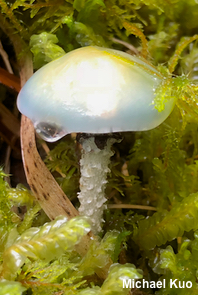
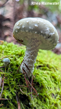
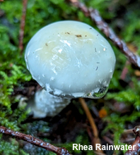
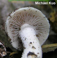
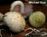
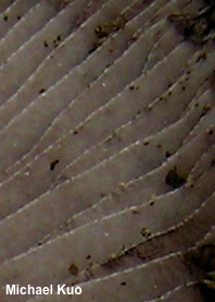
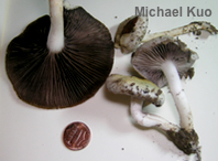
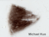
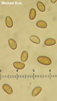
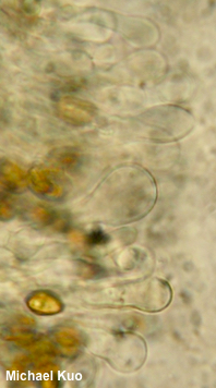
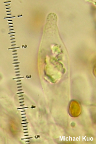
[ Basidiomycota > Agaricales > Strophariaceae > Stropharia. . . ] Stropharia aeruginosa by Michael Kuo, 20 April 2024 The original author of this gorgeous species (Curtis 1774) wrote that the mushroom is the color of verdigris when young and fresh, but that, "alas! . . . it loses that verdigris green, which on its first appearance renders it so conspicuous, the cap being often found of a pale yellowish brown color." The dramatic color change in Stropharia aeruginosa is reminiscent of the green-to-yellowish metamorphosis undergone by Gliophorus psittacinus, the "parrot mushroom." Unlike the parrot mushroom, however, Stropharia aeruginosa has purple-gray to purple-black gills, a purple-black spore print, and a sheathing ring on the stem (at least when young). Stropharia cyanea is nearly identical to the naked eye—although, in theory, its gill edges are not white and contrasting, like those of Stropharia aeruginosa; additionally its young stem is a bit less shaggy and its ring is less well developed. Under the microscope, however, Stropharia cyanea has very different gill edges; they are lined with fusiform chrysocystidia, whereas the gill edges of Stropharia aeruginosa are lined with non-refractive cystidia. Stropharia pseudocyanea (also known as "Stropharia albocyanea") is very similar, but it tends to grow in grassy areas and features a poorly developed ring; its cap is often much paler, when young, than the cap of Stropharia aeruginosa. Under the microscope the cheilocystidia of Stropharia pseudocyanea are narrower and less abruptly swollen. Thanks to Rhea Rainwater for documenting, collecting, and preserving Stropharia aeruginea for study; her collection is deposited in The Herbarium of Michael Kuo. Psilocybe aeruginosa is a synonym. Description: Ecology: Saprobic, growing alone or gregariously under hardwoods or conifers, on soil or on woody debris; summer and fall; originally described from Great Britain (Curtis 1774); widely distributed in Europe; in North America distributed in northern and montane areas, the Pacific Northwest, and the Midwest. The illustrated and described collections are from France, Arkansas, Illinois, and Washington. Cap: 3–8.5 cm; convex or broadly bell-shaped at first, becoming broadly convex, with or without a central bump—or nearly flat; very slimy when fresh; bald, or with scattered small scales, especially when young; when young deep blue-green, but soon fading to yellowish green or yellowish. Gills: Broadly attached to the stem but receding with maturity; close or nearly distant; short-gills frequent; whitish to pale gray at first, becoming purplish gray; edges white, contrasting with the faces at maturity. Stem: 3–8.5 cm long; 5–10 mm thick; equal; dry; when young with a sheathing ring with a flared and ragged upper edge, but with age sometimes merely featuring a ring zone; often with large, floccose, white scales when young; whitish; attached to white rhizomorphs. Flesh: White; unchanging when sliced. Odor and Taste: Odor not distinctive, or foul (almost reminiscent of the "green corn" odor found in some species of Inocybe); taste not distinctive, or somewhat radishlike. Chemical Reactions: KOH on cap surface negative to dull yellowish. Spore Print: Purplish black. Microscopic Features: Spores 7–10 x 4–5 µm; ellipsoid to subamygdaliform, with a tiny pore; smooth; yellow-brown in KOH. Basidia 24–28 x 5–6 µm; clavate; 4-sterigmate. Cheilocystidia 35–50 x 4–12 µm; capitate or subcapitate; smooth; thin-walled; hyaline in KOH. Cheilochrysocystidia not found. Pleurochrysocystidia 30–52 x 7–15 µm; fusoid-ventricose, with or without a small mucro—or ellipsoid; smooth; thin-walled; hyaline in KOH, with yellowish-refractive inclusions. Pileipellis an ixocutis; elements 3–10 µm wide, smooth, hyaline to yellowish in KOH; clamp connections present. REFERENCES: (W. Curtis, 1774) L. Quélet, 1872. (Kauffman, 1918; Stamets, 1978; Smith, Smith & Weber, 1979; Phillips, 1981; Arora, 1986; Watling & Gregory, 1987; Phillips, 1991/2005; Schalkwijk-Barendsen, 1991; Lincoff, 1992; Breitenbach & Kränzlin, 1995; Barron, 1999; Noordeloos, 1999; McNeil, 2006; Miller & Miller, 2006; Boccardo et al., 2008; Knudsen & Vesterholt, 2008; Trudell & Ammirati, 2009; Buczacki et al., 2013; Gminder & Böhning, 2017; Ryman, 2018; Læssøe & Petersen, 2019; Kibby, 2021; McKnight et al., 2021.) Herb. Kuo 09210602, 10261301, 10182301. This site contains no information about the edibility or toxicity of mushrooms. |
© MushroomExpert.Com |
|
Cite this page as: Kuo, M. (2024, April). Stropharia aeruginosa. Retrieved from the MushroomExpert.Com Web site: http://www.mushroomexpert.com/stropharia_aeruginosa.html |
