| Major Groups > Cup Fungi > Phylloscypha phyllogena |

|
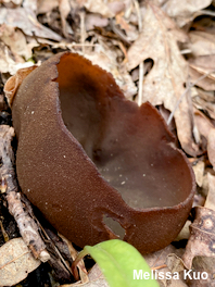
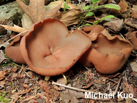
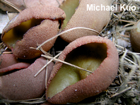
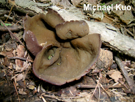
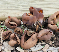
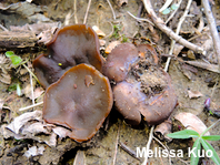
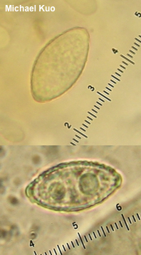
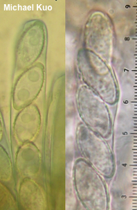
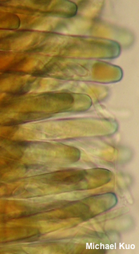
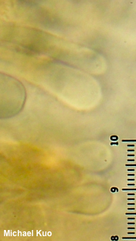
[ Ascomycota > Pezizales > Pezizaceae > Phylloscypha . . . ] Phylloscypha phyllogena by Michael Kuo, 13 August 2024 If you have found a large, brown cup fungus while hunting morels in the spring, odds are fairly high that it is Phylloscypha phyllogena—a commonly collected late spring to early summer species. However, plenty of other brown cups appear in morel season, from Disciotis venosa (very closely related to the morels) to cup-like species of Gyromitra (in the same genus as the false morels) to many others; microscopic analysis may be required to separate these mushrooms with certainty. Defining features for Phylloscypha phyllogena include its large size and vernal appearance, along with its subfusiform spores, which develop fine warts and ridges, along with fairly large "end caps." The cups are often reddish brown on the upper surface, but they are fairly variable in color; olive brown to yellowish brown specimens are also found regularly. Peziza phyllogena is a synomym, as is Peziza badioconfusa—and the latter name has often been used in field guides. However, after studying the 19th-century type collection of Peziza phyllogena, Pfister (1987) concluded it was the same as Peziza badioconfusa; since phyllogena is the older name, it takes precedence. Legaliana badia (formerly known as Peziza badia) is similar; however, it appears in late summer and fall, and its spores lack end caps but feature more developed ornamentation that is nearly reticulate. Description: Ecology: Probably mycorrhizal; growing gregariously or in small clusters, on the ground under hardwoods or conifers; sometimes, but not always, in woody debris; occasionally found in woodchips; spring and early summer; originally described from South Carolina (Cooke 1877); widely distributed in North America and in Europe. The illustrated and described collections are from Illinois, Michigan, and Pennsylvania. Fruiting Body: 2–12 cm across and 2–5 cm high; cup-shaped when young, often flattening to disc-shaped with age, or becoming irregular; when clustered often becoming pinched and contorted; upper surface dull and bald, usually a shade of brown—from reddish brown to purplish brown, olive brown, or yellow-brown; undersurface brown to reddish brown or purplish brown (sometimes lilac to purple near the point of attachment), roughened or finely mealy to granular with pustules, especially toward the margin; stem absent; basal mycelium white; attached to the substrate at a central location; flesh brown to whitish, brittle, unchanging when sliced. Odor: Not distinctive. Chemical Reactions: KOH negative on all surfaces; Melzer's reagent green to blackish green on upper surface. Spore Print: White. Microscopic Features: Spores 17–22 x 7–10 µm including ornamentation; subfusiform to fusiform at maturity; at first smooth, but soon becoming verruculose to verrucose with fine isolated warts 0.25–0.5 µm high; often developing broad, flattened, apical caps; 1- or 2-guttulate, especially during development; hyaline in KOH; walls cyanophilic. Asci 8-spored; with amyloid tips. Paraphyses hardly exceeding the asci; filiform; 2–5 µm wide at the apex; apices subclavate or merely rounded; smooth; hyaline in KOH. REFERENCES: (M. C. Cooke, 1877) N. van Vooren, 2020. (Seaver, 1942; Korf, 1954; Wells & Kempton, 1967; Elliott & Kaufert, 1973; Smith, Smith & Weber, 1981; Pfister, 1987; Phillips, 1991/2005; Lincoff, 1992; Roody, 2003; McNeil, 2006; Beug et al., 2012; Raymundo et al., 2012; Medel et al., 2013; Kuo & Methven, 2014; Elliott & Stephenson, 2018; Kajevska et al., 2018; van Vooren, 2020; McKnight et al., 2021; van Vooren et al., 2021.) Herb. Kuo 05080302, 05040602, 05120605, 05060701, 05040801, 05250801, 05021101, 04231602, 05131601, 06081601, 04091701, 05032001. This site contains no information about the edibility or toxicity of mushrooms. |
© MushroomExpert.Com |
|
Cite this page as: Kuo, M. (2024, August). Phylloscypha phyllogena. Retrieved from the MushroomExpert.Com Web site: http://www.mushroomexpert.com/phylloscypha_phyllogena.html. |
