| Major Groups > Polypores > Ganoderma > Ganoderma tsugae |

|
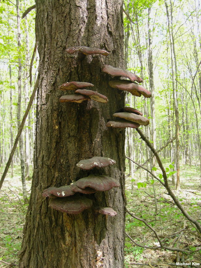
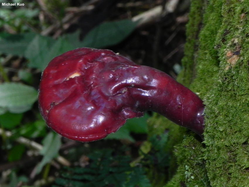
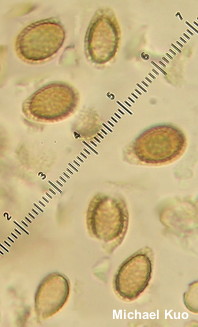
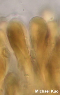
Ganoderma tsugae [ Basidiomycota > Polyporales > Polyporaceae > Ganoderma . . . ] by Michael Kuo This incredible mushroom is found almost exclusively on the wood of eastern hemlocks, and its range parallels that of the tree: northern and montane areas east of the Rocky Mountains. It is one of the "laccate" species of Ganoderma, which means it features a beautifully lacquered-looking cap surface. Like other species of Ganoderma its pore surface bruises brown and its spores have double walls with a chambered appearance. Ganoderma oregonense is a similar species found in the Pacific Northwest and California on the wood of various conifers; it features larger spores. Until recently Ganoderma oregonense and Ganoderma tsugae were thought to coexist in the Pacific Northwest, but recent study of hundreds of North American Ganoderma collections by Loyd and collaborators (2017, 2018) failed to support this idea. Description: Ecology: Parasitic and saprobic on the wood of eastern hemlock and perhaps other conifers; causing a white rot (usually a butt rot) of the heartwood; growing alone, scattered, or gregariously; annual; spring through fall; distributed in the northern Midwest, northeastern North America, and the Appalachian Mountains (where eastern hemlocks occur). The illustrated and described collections are from Michigan and Ohio. Cap: 4–16 cm; at first irregularly knobby or elongated, but by maturity more or less fan- or kidney-shaped; with a shiny, varnished surface often roughly arranged into lumpy "zones"; bald; dark red to orangish red or reddish brown when mature; when young often with zones of bright yellow and white toward the margin. Pore Surface: Whitish, becoming dingy reddish brown in age; usually bruising brown; with 4–6 tiny (nearly invisible to the naked eye) circular pores per mm; tubes to 1 cm deep. Stem: Sometimes absent, but more commonly present; 3–14 cm long; up to 3 cm thick; equal or irregular; varnished and colored like the cap; often distinctively angled away from one side of the cap. Flesh: Whitish when fresh; fairly soft when young, but soon tougher; concentric growth zones and melanoid bands (see discussion) absent. Chemical Reactions: KOH instantly black on flesh and tubes. Spore Print: Brown. Microscopic Features: Spores 8–12 x 5–7 µm; including the hyaline vesicular appendix; more or less ellipsoid, with a truncated end; appearing double-walled, with a series of "pillars" between the walls; finely stippled; inamyloid; brown in KOH. Cystidia and setae not found. Hyphal system trimitic. Clamp connections present. Terminal cells on cap surface clavate; 7.5–12.5 µm wide; thick-walled; golden in KOH. REFERENCES: Murrill, 1902. (Overholts, 1953; Smith, Smith & Weber, 1981; Gilbertson & Ryvarden, 1986; Phillips, 1991/2005; Lincoff, 1992; Roody, 2003; McNeil, 2006; Kuo & Methven, 2014; Woehrel & Light, 2017; Baroni, 2017; Loyd et al., 2017; Elliott & Stephenson, 2018; Loyd et al., 2018.) Herb. Kuo 09110402, 07271201. This website contains no information about the edibility or toxicity of mushrooms. |
© MushroomExpert.Com |

|
Cite this page as: Kuo, M. (2019, January). Ganoderma tsugae. Retrieved from the MushroomExpert.Com Web site: http://www.mushroomexpert.com/ganoderma_tsugae.html |