| Major Groups > Gilled Mushrooms > Dark-Spored > Conocybe & Pholiotina > Conocybe siennophylla |

|
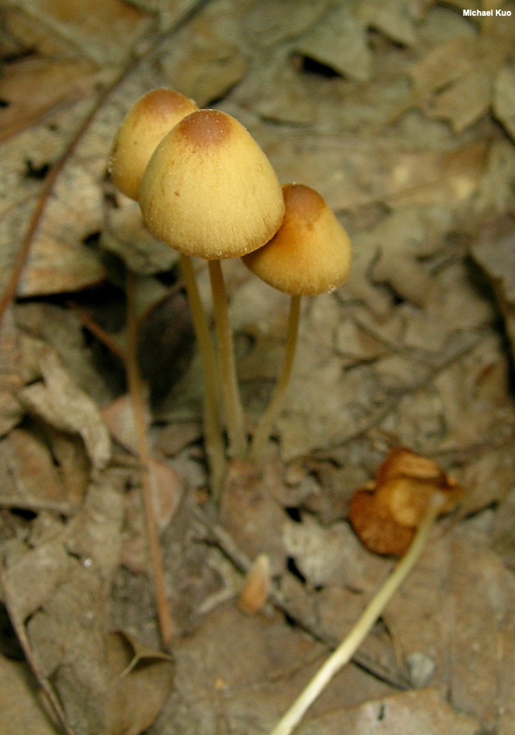
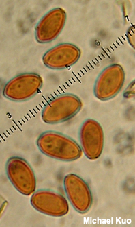
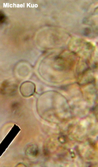
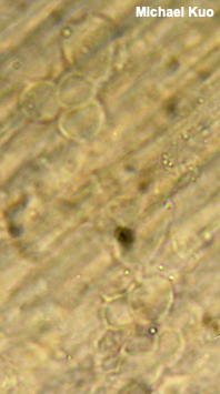
[ Basidiomycota > Agaricales > Bolbitiaceae > Conocybe . . . ] Conocybe siennophylla by Michael Kuo, January 2019 Defining features for Conocybe siennophylla include the yellowish-brownish cap, spore dimensions (see below), and the anatomy of the cystidia on the stem surface, which are not shaped like bowling pins (the cystidia on the gills' edges, however, are shaped like bowling pins). If painstaking microscopy does not interest you, I will certainly not blame you if you take a bye on Conocybe identification. Conocybe velutipes is virtually identical to the naked eye, but features larger spores with thicker walls. Description: Ecology: Saprobic; growing alone, scattered, or gregariously in woods or in grassy areas; often found in disturbed ground; late spring through fall; North American distribution uncertain, but reported from the Midwest and the West Coast in online records of major herbaria. The illustrated and described collection is from Illinois. Cap: 0.5–2 cm; conical to bell-shaped when young, becoming broadly bell-shaped or convex; dry; becoming finely lined for 2–3 mm; yellow-brown, fading markedly to yellowish tan, but retaining a darker center. Gills: Attached to the stem; close or crowded; colored like the cap at first, becoming cinnamon; dissolving in hot weather. Stem: 4–8 cm long; 1–2 mm thick; fragile; more or less equal; whitish to yellowish at first, becoming brownish; bald or finely powdery-fibrillose; basal mycelium white. Flesh: Insubstantial. Odor and Taste: Not distinctive. Spore Print: Cinnamon brown or reddish brown. Chemical Reactions: KOH dull red on cap surface. Microscopic Features: Spores 9–11 x 5–6 µm; ellipsoid, with a pore about 1 µm across; walls about 0.5 µm thick; smooth; orangish brown in KOH. Basidia 4-sterigmate. Brachybasidioles present. Pleurocystidia not found. Cheilocystidia 17–25 x 7–12 µm; lecythiform, with a severely narrowed neck between a clavate to ellipsoid bottom portion and a subglobose head; thin-walled; smooth; hyaline in KOH. Pileipellis hymeniform/cellular with subglobose terminal cells 10–25 µm across; hyaline to brownish in KOH. Caulocystidia in bundles; 5–10 x 4–6 µm; subglobose to ellipsoid or sublageniform; smooth; hyaline in KOH. REFERENCES: (Berkeley & Broome, 1871) Singer ex Chiari & Papetti, 2016. (Saccardo, 1887; Phillips, 1981; Watling, 1982; Breitenbach & Kränzlin, 1995; Arnolds, 2005; Hausknecht & Vesterholt, 2018.) Herb. Kuo 05100701. This site contains no information about the edibility or toxicity of mushrooms. |
© MushroomExpert.Com |
|
Cite this page as: Kuo, M. (2019, January). Conocybe siennophylla. Retrieved from the MushroomExpert.Com Web site: http://www.mushroomexpert.com/conocybe_siennophylla.html |